Author: Cornelius Onye Nichodemus
Introduction
Several variations exist in living organisms both phenotypic and genotypic. These variations are usually as a result of the genomic (DNA and RNA) makeup of each individual which makes living organisms differ from one another in size, structure, function etc. In order to study this genomic make up of living organisms, electrophoresis techniques is applied for separation and analysis of macromolecules based on their size and charge.
GEL ELECTROPHORESIS
Is a technique used for separation and analysis of macromolecules (DNA, RNA and Proteins) and their fragments, based on their size and charge. It is used in biochemistry and molecular biology to separate a mixed population of DNA and RNA fragments by length, to estimate the size of DNA and RNA fragments or to separate proteins by charge or size. Electrophoresis techniques is based on the fact that nucleic acid molecules (DNA, RNA) is separated by applying electric field to move the negatively charged molecules towards the positively charged anode. Shorter molecules move faster or migrate further than longer ones because shorter molecules migrate more easily through the pores of the gel. This process is called sieving. Proteins are separated by charge in agarose because the pores of the gel are too charged to sieve proteins. Gel electrophoresis can also be separation of nanoparticles.
DNA Gel electrophoresis is usually performed for analytical purposes often after amplification of DNA via polymerase chain reaction (PCR), but may be used as a preparatory technique prior to the use of other methods such as mass spectrometry, restriction fragments length polymorphism (RFLP), PCR, cloning, DNA sequencing or Southern blotting.
TYPES OF GEL
There are basically two types of gels used and they are;
1. Agarose gel
2. Polyacrylamide gel
Each type of gel is well suited to different types and sizes of analyze.
1. AGAROSE GELS: they are made from natural polysaccharide polymers extracted from seaweed.
- have low resolving power for DNA but have great range of separation and are therefore used for DNA fragments of usually 50-20000bp in size
- a resolution of over 6Mb is possible with pulsed field gel electrophoresis (PFGE)
- agarose gel run horizontally and do not have uniform pore size
- the casting methodology is thermal and easy
- gel setting is physical
2. POLYACRYLAMIDE GEL (PAGE): they are used for protein analysis
- have a resolving power for small fragments of DNA (5-500bp)
- run in a vertical configuration
- forms a chemical polymerisation reaction
- have uniform pore size
MODE OF OPERATION
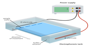
Figure 1. Illustration of DNA electrophoresis equipment used to separate DNA fragments by size. A gel sits within a tank of buffer. The DNA samples are placed in Wells at one end of the gel and an electric current is passed across the gel. The negatively charged DNA moves towards the positively charged anode.
Source: Genome Research Limited
Basically, electrophoresis is a process which enables the sorting of molecules based on their size. Using an electric field, molecules (DNA) can be made to move through a gel made of agar or polyacrylamide. The electric field consists of a negative charge at one end which pushes the molecules through the gel and a positive charge at the other end that pulls the molecules through the gel. The molecules being sorted are dispensed into a well in the gel material. The gel is placed in an electrophoresis chamber and connected to a power source. When the electric current is applied, the longer molecules move more slowly through the gel while the smaller molecules move faster. The different sized molecules form distinct bands on the gel as shown in figure 2.
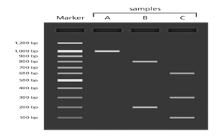
Figure 2. Illustration showing DNA bands separated on a gel using the UV transilluminator. The length of the DNA fragments is compared to a marker containing fragments of known lengths.
Source: Genome Research Limited
VISUALIZATION
When the fragments have been separated, examine the hell and see what sizes of bands that are found on it. The genus stained with a DNA binding dye we'd placed under UV light, this enables the different DNA fragments to be seen at various locations of the gel.
Alternatively, gels are visualized by using UV transilluminator by staining with ethidium bromide. Each gel is run along with DNA molecular weight marker of known molecular size that are used as a standard to determine the size of the unknown fragments.
Bands are well defined line of DNA on a gel. Each band contains a large number of DNA fragments of the same size that have migrated as a group to the same location.
APPLICATIONS
1. Analysis of PCR products e.g molecular genetic diagnosis or genetic fingerprinting
2. Separation of restricted genomic DNA prior to southern transfer or of RNA to northern transfer
3. Estimation of the use of DNA molecules following enzymes digestion e.g in restriction mapping of cloned DNA.
Furthermore, gel electrophoresis is used in molecular biology, forensics, genetics, microbiology and biochemistry.
In conclusion, gel electrophoresis has enabled an advance study of macromolecules (DNA, RNA and proteins) through electric current which is now applied in PCR analysis, separation of restricted genomic DNA etc.
REFERENCES
- Biotechniques laboratory electrophoresis demonstration from the University of Utah's Genetic Science Learning Center.
- Milan Bier (ed.) (1959). Electrophoresis. Theory, Methods and Applications (3rd printing ed.). Academic Press. p. 225. OCLC 1175404. LCC 59-7676.
- www.khanacademy.org/gel-electrophoresis
About Author / Additional Info:
I am a First Class graduate of Plant Science and Biotechnology from University of port Harcourt.